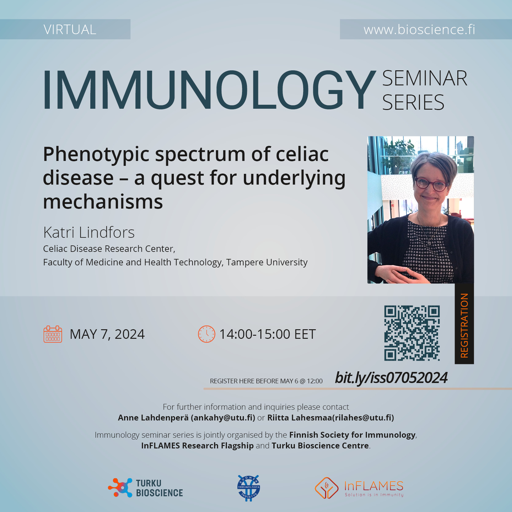
Immunology Seminar Series, Katri Lindfors
May 7th online at 14 – 15 (Finland time)
Virtual event
Katri Lindfors, Celiac Disease Research Center, Faculty of Medicine and Health Technology, Tampere University: Phenotypic spectrum of celiac disease – a quest for underlying mechanisms
Please register latest May 6th at https://link.webropolsurveys.com/S/EE67FF858AC0A584
Host: Riitta Lahesmaa ( rilahes@utu.fi )
Immunology seminar series is jointly organised by the Finnish Society for Immunology, InFLAMES Flagship and Turku Bioscience. For further information contact Anne Lahdenperä ( ankahy@utu.fi ) or Riitta Lahesmaa ( rilahes@utu.fi ), University of Turku.
***
Katri Lindfors is a professor of molecular biology and the vice director of Celiac Disease Research Center at Tampere University. She is a member of the organizing committee of the Tampere Celiac Disease Symposium, the scientific advisory board of the patient organization Finnish Celiac Society and International Society for the Study of Celiac Disease.
She has conducted translational celiac disease research over 20 years and published over 100 peer-reviewed articles. Her previous work has contributed to the understanding of factors involved in celiac disease pathogenesis including the potential role of enteroviruses as a triggering factor and the biological functions of celiac disease specific autoantibodies targeting a self-protein transglutaminase 2 (TG2). Currently her group focuses on identifying factors contributing to the heterogenous clinical picture of celiac disease using dermatitis herpetiformis, the skin manifestation of celiac disease, as a model phenotype. Up to now, her results indicate that in addition to a distinct genetic predisposition, an autoimmune response against a TG3, a homologue of celiac disease autoantigen TG2 is implicated in dermatitis herpetiformis. Ongoing studies are addressing the questions whether dermatitis herpetiformis pathogenesis is initiated in the small bowel mucosa and how the disease spreads from gut to skin as a consequence of unrecognized and untreated celiac disease.
References:
- Das S, Stamnaes J, Kemppainen E, Hervonen K, Lundin KEA, Parmar N, Jahnsen FL, Jahnsen J, Lindfors K, Salmi T, Iversen R, Sollid LM. Separate Gut Plasma Cell Populations Produce Auto-Antibodies against Transglutaminase 2 and Transglutaminase 3 in Dermatitis Herpetiformis. Adv Sci (Weinh) 2023;10:e2300401. doi: 10.1002/advs.202300401.
- Kalliokoski S, Mansikka E, de Kauwe A, Huhtala H, Saavalainen P, Kurppa K, Hervonen K, Reunala T, Kaukinen K, Salmi T, Lindfors K. Gliadin-induced ex vivo T cell response in dermatitis herpetiformis: A predictor of clinical relapse on gluten challenge? J Invest Dermatol, 2020;140:1867-1869.e2 doi: 10.1016/j.jid.2019.12.038.
- Lindfors K, Ciacci C, Kurppa K, Lundin K, Makharia G, Mearin ML, Murray J, Verdu E, Kaukinen K. Coeliac Disease. Nature Reviews Disease Primers 2019;5:3 DOI:10.1038/s41572-018-0054-z.
- Hietikko M, Hervonen K, Salmi T, Ilus T, Zone JJ, Kaukinen K, Reunala T, Lindfors K. Disappearance of epidermal transglutaminase and IgA deposits from the papillary dermis of dermatitis herpetiformis patients after a long-term gluten-free diet. Brit J Dermatol 2018;178:e198-e201. doi: 10.1111/bjd.15995.

